Pelvic Ultrasound and Transvaginal Ultrasound: Key Clinical Differences
For women experiencing pelvic pressure, irregular menstrual cycles, abnormal spotting, or unexplained lower abdominal discomfort, ultrasound is universally regarded as the gold standard first line diagnostic tool. Safe, gentle, and 100% radiation free, ultrasound visualizes the reproductive organs with notable detail. Often, patients wonder about the specific difference between a transabdominal pelvic ultrasound and an endovaginal scan, and why physicians frequently recommend combining both methods.
The Transabdominal Pelvic Scan: Broad Overview
A transabdominal pelvic ultrasound is performed externally. The sonographer applies warm acoustic gel across your lower abdomen and sweeps the transducer above the pubic bone. This approach provides an expansive panoramic view of the entire lower pelvis, mapping the position of the uterus, ovaries, and adjacent structures.
Because the pelvic bones and intestines can obscure acoustic waves, a transabdominal exam requires a full urinary bladder. Drinking thirty two ounces of water approximately one hour before your appointment fills the bladder, gently pushing the intestines upward and serving as an acoustic liquid window that transmits sound waves deep into the pelvic cavity.
The Transvaginal Ultrasound: High Resolution Precision
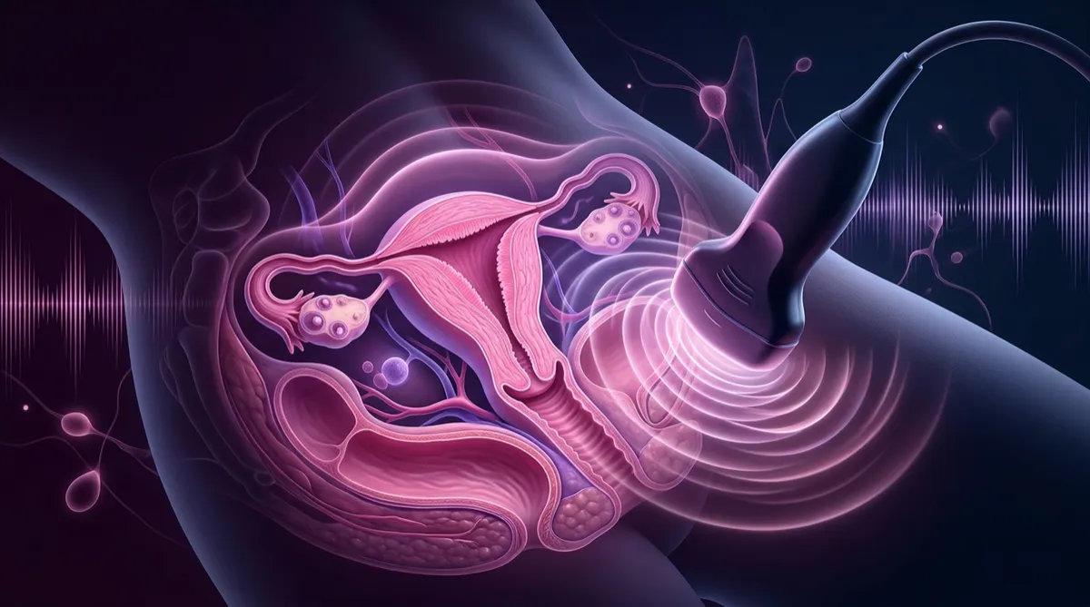
While an external scan provides broad structural orientation, a transvaginal ultrasound offers exceptional close up resolution. By using a slender, specialized transducer covered in a sterile protective sheath and warm gel, the imaging probe rests close to the pelvic organs. This proximity bypasses abdominal wall tissue, yielding sharp microscopic detail of the endometrial lining, uterine muscular wall, and ovarian follicles.
Before beginning a transvaginal scan, patients completely empty their bladder. Our registered female sonographers prioritize dignity, privacy, and gentle pacing throughout the entire appointment, ensuring you remain informed and comfortable during every moment.
What Each Examination Identifies
When interpreted together by Board Certified Radiologists, these complementary imaging approaches provide diagnostic clarity across several common women health conditions:
- Ovarian Cysts and Follicles: Differentiating between simple fluid filled physiological cysts and complex solid lesions or endometriomas.
- Uterine Fibroids and Polyps: Locating benign muscular growths (fibroids) within the uterine wall and identifying endometrial polyps that cause heavy bleeding.
- Endometrial Thickness: Accurately measuring the mucosal lining of the uterus to evaluate postmenopausal bleeding or hormonal irregularities.
- Early Pregnancy Viability: Confirming intrauterine gestational sacs, heartbeat detection, and accurate dating during the first trimester.
Compassionate Outpatient Care in Chicago
Undergoing sensitive pelvic imaging should never feel clinical, rushed, or stressful. At Perfect Diagnostic Ultrasound, patients receive personal, respectful care in a peaceful setting with experienced sonographers and rapid twenty four to forty eight hour radiologist reports.
Medical Disclaimer: This article is designed solely for informational and educational guidance. It is not a substitute for professional clinical diagnosis or emergency medical care. If you experience sudden, severe unilateral pelvic pain accompanied by dizziness or heavy hemorrhage, contact emergency medical services immediately.
